Profile

Affiliate Associate Professor.
Develops optical tools to probe biology at the border of physics and chemistry.
Publications
Malyshev, Dmitry; Öberg, Rasmus; Landström, Lars; Andersson, Per Ola; Dahlberg, Tobias; Andersson, Magnus
pH-induced changes in Raman, UV–vis absorbance, and fluorescence spectra of dipicolinic acid (DPA) Journal Article
In: Spectrochimica Acta Part A: Molecular and Biomolecular Spectroscopy, vol. 271, pp. 120869, 2022, ISSN: 13861425.
@article{Malyshev2022a,
title = {pH-induced changes in Raman, UV–vis absorbance, and fluorescence spectra of dipicolinic acid (DPA)},
author = {Dmitry Malyshev and Rasmus Öberg and Lars Landström and Per Ola Andersson and Tobias Dahlberg and Magnus Andersson},
url = {https://linkinghub.elsevier.com/retrieve/pii/S1386142522000178},
doi = {10.1016/j.saa.2022.120869},
issn = {13861425},
year = {2022},
date = {2022-04-01},
journal = {Spectrochimica Acta Part A: Molecular and Biomolecular Spectroscopy},
volume = {271},
pages = {120869},
keywords = {},
pubstate = {published},
tppubtype = {article}
}
Malyshev, Dmitry; Öberg, Rasmus; Dahlberg, Tobias; Wiklund, Krister; Landström, Lars; Andersson, Per Ola; Andersson, Magnus
Laser induced degradation of bacterial spores during micro-Raman spectroscopy Journal Article
In: Spectrochimica Acta Part A: Molecular and Biomolecular Spectroscopy, vol. 265, pp. 120381, 2022, ISSN: 13861425.
@article{Malyshev2022,
title = {Laser induced degradation of bacterial spores during micro-Raman spectroscopy},
author = {Dmitry Malyshev and Rasmus Öberg and Tobias Dahlberg and Krister Wiklund and Lars Landström and Per Ola Andersson and Magnus Andersson},
url = {https://doi.org/10.1016/j.saa.2021.120381 https://linkinghub.elsevier.com/retrieve/pii/S1386142521009586},
doi = {10.1016/j.saa.2021.120381},
issn = {13861425},
year = {2022},
date = {2022-01-01},
journal = {Spectrochimica Acta Part A: Molecular and Biomolecular Spectroscopy},
volume = {265},
pages = {120381},
publisher = {The Author(s)},
keywords = {},
pubstate = {published},
tppubtype = {article}
}
Panadeiro, Veronica; Rodriguez, Alvaro; Henry, Jason; Wlodkowic, Donald; Andersson, Magnus
A review of 28 free animal-tracking software applications: current features and limitations Journal Article
In: Lab Animal, vol. 50, no. 9, pp. 246–254, 2021, ISSN: 0093-7355.
@article{Panadeiro2021,
title = {A review of 28 free animal-tracking software applications: current features and limitations},
author = {Veronica Panadeiro and Alvaro Rodriguez and Jason Henry and Donald Wlodkowic and Magnus Andersson},
url = {http://dx.doi.org/10.1038/s41684-021-00811-1 https://www.nature.com/articles/s41684-021-00811-1},
doi = {10.1038/s41684-021-00811-1},
issn = {0093-7355},
year = {2021},
date = {2021-09-01},
journal = {Lab Animal},
volume = {50},
number = {9},
pages = {246--254},
publisher = {Springer US},
abstract = {Well-quantified laboratory studies can provide a fundamental understanding of animal behavior in ecology, ethology and ecotoxicology research. These types of studies require observation and tracking of each animal in well-controlled and defined arenas, often for long timescales. Thus, these experiments produce long time series and a vast amount of data that require the use of software applications to automate the analysis and reduce manual annotation. In this review, we examine 28 free software applications for animal tracking to guide researchers in selecting the software that might best suit a particular experiment. We also review the algorithms in the tracking pipeline of the applications, explain how specific techniques can fit different experiments, and finally, expose each approach's weaknesses and strengths. Our in-depth review includes last update, type of platform, user-friendliness, off- or online video acquisition, calibration method, background subtraction and segmentation method, species, multiple arenas, multiple animals, identity preservation, manual identity correction, data analysis and extra features. We found, for example, that out of 28 programs, only 3 include a calibration algorithm to reduce image distortion and perspective problems that affect accuracy and can result in substantial errors when analyzing trajectories and extracting mobility or explored distance. In addition, only 4 programs can directly export in-depth tracking and analysis metrics, only 5 are suited for tracking multiple unmarked animals for more than a few seconds and only 11 have been updated in the period 2019–2021.},
keywords = {},
pubstate = {published},
tppubtype = {article}
}
Näsström, Thomas; Dahlberg, Tobias; Malyshev, Dmitry; Ådén, Jörgen; Andersson, Per Ola; Andersson, Magnus; Karlsson, Björn C G
Synthetic NAC 71-82 Peptides Designed to Produce Fibrils with Different Protofilament Interface Contacts Journal Article
In: International Journal of Molecular Sciences, vol. 22, no. 17, pp. 9334, 2021, ISSN: 1422-0067.
@article{Nasstrom2021,
title = {Synthetic NAC 71-82 Peptides Designed to Produce Fibrils with Different Protofilament Interface Contacts},
author = {Thomas Näsström and Tobias Dahlberg and Dmitry Malyshev and Jörgen Ådén and Per Ola Andersson and Magnus Andersson and Björn C G Karlsson},
url = {https://www.mdpi.com/1422-0067/22/17/9334},
doi = {10.3390/ijms22179334},
issn = {1422-0067},
year = {2021},
date = {2021-08-01},
journal = {International Journal of Molecular Sciences},
volume = {22},
number = {17},
pages = {9334},
abstract = {Alpha-synucleinopathies are featured by fibrillar inclusions in brain cells. Although $alpha$-synuclein fibrils display structural diversity, the origin of this diversity is not fully understood. We used molecular dynamics simulations to design synthetic peptides, based on the NAC 71-82 amino acid fragment of $alpha$-synuclein, that govern protofilament contacts and generation of twisted fibrillar polymorphs. Four peptides with structures based on either single or double fragments and capped or non-capped ends were selected for further analysis. We determined the fibrillar yield and the structures from these peptides found in the solution after fibrillisation using protein concentration determination assay and circular dichroism spectroscopy. In addition, we characterised secondary structures formed by individual fibrillar complexes using laser-tweezers Raman spectroscopy. Results suggest less mature fibrils, based on the lower relative $beta$-sheet content for double- than single-fragment peptide fibrils. We confirmed this structural difference by TEM analysis which revealed, in addition to short protofibrils, more elongated, twisted and rod-like fibril structures in non-capped and capped double-fragment peptide systems, respectively. Finally, time-correlated single-photon counting demonstrated a difference in the Thioflavin T fluorescence lifetime profiles upon fibril binding. It could be proposed that this difference originated from morphological differences in the fibril samples. Altogether, these results highlight the potential of using peptide models for the generation of fibrils that share morphological features relevant for disease, e.g., twisted and rod-like polymorphs.},
keywords = {},
pubstate = {published},
tppubtype = {article}
}
Dahlberg, Tobias; Andersson, Magnus
Optical design for laser tweezers Raman spectroscopy setups for increased sensitivity and flexible spatial detection Journal Article
In: Applied Optics, vol. 60, no. 16, pp. 4519, 2021, ISSN: 1559-128X.
@article{Dahlberg2021,
title = {Optical design for laser tweezers Raman spectroscopy setups for increased sensitivity and flexible spatial detection},
author = {Tobias Dahlberg and Magnus Andersson},
url = {https://www.osapublishing.org/abstract.cfm?URI=ao-60-16-4519},
doi = {10.1364/AO.424595},
issn = {1559-128X},
year = {2021},
date = {2021-06-01},
journal = {Applied Optics},
volume = {60},
number = {16},
pages = {4519},
abstract = {We demonstrate a method to double the collection efficiency in Laser Tweezers Raman Spectroscopy (LTRS) by collecting both the forward and back-scattered light in a single-shot multitrack measurement. Our method can collect signals at different sample volumes, granting both the pinpoint spatial selectivity of confocal Raman and the bulk sensitivity of non-confocal Raman simultaneously. Further, we display that our approach allows for reduced detector integration time and laser power. Thus, our method will enable the monitoring of biological samples sensitive to high intensities for longer times. Additionally, we demonstrate that by a simple modification, we can add polarization sensitivity and retrieve extra biochemical information.},
keywords = {},
pubstate = {published},
tppubtype = {article}
}
Nilsson, Daniel P. G.; Dahlberg, Tobias; Andersson, Magnus
Step-by-step guide to 3D print motorized rotation mounts for optical applications Journal Article
In: Applied Optics, vol. 60, no. 13, pp. 3764, 2021, ISSN: 1559-128X.
@article{Nilsson2021,
title = {Step-by-step guide to 3D print motorized rotation mounts for optical applications},
author = {Daniel P. G. Nilsson and Tobias Dahlberg and Magnus Andersson},
url = {https://www.osapublishing.org/abstract.cfm?URI=ao-60-13-3764},
doi = {10.1364/AO.422695},
issn = {1559-128X},
year = {2021},
date = {2021-05-01},
journal = {Applied Optics},
volume = {60},
number = {13},
pages = {3764},
abstract = {Motorized rotation mounts and stages are versatile instruments that introduce computer control to optical systems, enabling automation and scanning actions. They can be used for intensity control and position adjustments, etc. However, these rotation mounts come with a hefty price tag, and this limits their use. This work shows how to build two different types of motorized rotation mounts for 1" optics, using a 3D printer and off-the-shelf components. The first is intended for reflective elements, like mirrors and gratings, and the second for transmissive elements, like polarizers and retarders. We evaluate and compare their performance to commercial systems based on velocity, resolution, accuracy, backlash, and axis wobble. Also, we investigate the angular stability using Allan variance analysis. The results show that our mounts perform similar to systems costing more than 2000 Euro, while also being quick to build and costing less than 200 Euro. As a proof of concept, we show how to control lasers used in an optical tweezers and Raman spectroscopy setup. When used for this, the 3D printed motorized rotational mounts provide intensity control with a resolution of $0.03$ percentage points or better.},
keywords = {},
pubstate = {published},
tppubtype = {article}
}
Baker, Joseph L; Dahlberg, Tobias; Bullitt, Esther; Andersson, Magnus
Impact of an alpha helix and a cysteine–cysteine disulfide bond on the resistance of bacterial adhesion pili to stress Journal Article
In: Proceedings of the National Academy of Sciences, vol. 118, no. 21, pp. e2023595118, 2021, ISSN: 0027-8424.
@article{Baker2021,
title = {Impact of an alpha helix and a cysteine–cysteine disulfide bond on the resistance of bacterial adhesion pili to stress},
author = {Joseph L Baker and Tobias Dahlberg and Esther Bullitt and Magnus Andersson},
url = {http://biorxiv.org/content/early/2021/01/19/2021.01.18.427124.abstract http://www.pnas.org/lookup/doi/10.1073/pnas.2023595118},
doi = {10.1073/pnas.2023595118},
issn = {0027-8424},
year = {2021},
date = {2021-05-01},
journal = {Proceedings of the National Academy of Sciences},
volume = {118},
number = {21},
pages = {e2023595118},
abstract = {Escherichia coli express adhesion pili that mediate attachment to host cell surfaces and are exposed to body fluids in the urinary and gastrointestinal tracts. Pilin subunits are organized into helical polymers, with a tip adhesin for specific host binding. Pili can elastically unwind when exposed to fluid flow forces, reducing the adhesin load, thereby facilitating sustained attachment. Here we investigate biophysical and structural differences of pili commonly expressed on bacteria that inhabit the urinary and intestinal tracts. Optical tweezers measurements reveal that class 1a pili of uropathogenic E. coli (UPEC), as well as class 1b of enterotoxigenic E. coli (ETEC), undergo an additional conformational change beyond pilus unwinding, providing significantly more elasticity to their structure than ETEC class 5 pili. Examining structural and steered molecular dynamics simulation data, we find that this difference in class 1 pili subunit behavior originates from an $alpha$-helical motif that can unfold when exposed to force. A disulfide bond cross-linking $beta$-strands in class 1 pili stabilizes subunits, allowing them to tolerate higher forces than class 5 pili that lack this covalent bond. We suggest that these extra contributions to pilus resiliency are relevant for the UPEC niche, since resident bacteria are exposed to stronger, more transient drag forces compared to those experienced by ETEC bacteria in the mucosa of the intestinal tract. Interestingly, class 1b ETEC pili include the same structural features seen in UPEC pili, while requiring lower unwinding forces that are more similar to those of class 5 ETEC pili.},
keywords = {},
pubstate = {published},
tppubtype = {article}
}
Malyshev, Dmitry; Dahlberg, Tobias; Wiklund, Krister; Andersson, Per Ola; Henriksson, Sara; Andersson, Magnus
Mode of Action of Disinfection Chemicals on the Bacterial Spore Structure and Their Raman Spectra Journal Article
In: Analytical Chemistry, vol. 93, no. 6, pp. 3146–3153, 2021, ISSN: 0003-2700.
@article{Malyshev2021,
title = {Mode of Action of Disinfection Chemicals on the Bacterial Spore Structure and Their Raman Spectra},
author = {Dmitry Malyshev and Tobias Dahlberg and Krister Wiklund and Per Ola Andersson and Sara Henriksson and Magnus Andersson},
url = {https://pubs.acs.org/doi/10.1021/acs.analchem.0c04519},
doi = {10.1021/acs.analchem.0c04519},
issn = {0003-2700},
year = {2021},
date = {2021-02-01},
journal = {Analytical Chemistry},
volume = {93},
number = {6},
pages = {3146--3153},
publisher = {American Chemical Society},
keywords = {},
pubstate = {published},
tppubtype = {article}
}
Enevold, Jenny; Dahlberg, Tobias; Stangner, Tim; Tang, Shi; Lindh, E. Mattias; Gracia-Espino, Eduardo; Andersson, Magnus; Edman, Ludvig
Tunable Two-Dimensional Patterning of a Semiconducting and Nanometer-Thin C 60 Fullerene Film Using a Spatial Light Modulator Journal Article
In: ACS Applied Nano Materials, pp. acsanm.0c00793, 2020, ISSN: 2574-0970.
@article{Enevold2020,
title = {Tunable Two-Dimensional Patterning of a Semiconducting and Nanometer-Thin C 60 Fullerene Film Using a Spatial Light Modulator},
author = {Jenny Enevold and Tobias Dahlberg and Tim Stangner and Shi Tang and E. Mattias Lindh and Eduardo Gracia-Espino and Magnus Andersson and Ludvig Edman},
url = {https://pubs.acs.org/doi/10.1021/acsanm.0c00793},
doi = {10.1021/acsanm.0c00793},
issn = {2574-0970},
year = {2020},
date = {2020-05-01},
journal = {ACS Applied Nano Materials},
pages = {acsanm.0c00793},
keywords = {},
pubstate = {published},
tppubtype = {article}
}
Escamez, Sacha; André, Domenique; Sztojka, Bernadette; Bollhöner, Benjamin; Hall, Hardy; Berthet, Béatrice; Voß, Ute; Lers, Amnon; Maizel, Alexis; Andersson, Magnus; Bennett, Malcolm; Tuominen, Hannele
Cell Death in Cells Overlying Lateral Root Primordia Facilitates Organ Growth in Arabidopsis Journal Article
In: Current Biology, vol. 30, no. 3, pp. 455–464.e7, 2020, ISSN: 09609822.
@article{Escamez2020,
title = {Cell Death in Cells Overlying Lateral Root Primordia Facilitates Organ Growth in Arabidopsis},
author = {Sacha Escamez and Domenique André and Bernadette Sztojka and Benjamin Bollhöner and Hardy Hall and Béatrice Berthet and Ute Voß and Amnon Lers and Alexis Maizel and Magnus Andersson and Malcolm Bennett and Hannele Tuominen},
url = {https://linkinghub.elsevier.com/retrieve/pii/S0960982219315805},
doi = {10.1016/j.cub.2019.11.078},
issn = {09609822},
year = {2020},
date = {2020-02-01},
journal = {Current Biology},
volume = {30},
number = {3},
pages = {455--464.e7},
abstract = {Unlike animal development, plant organ growth is widely accepted to be determined by cell division without any contribution of cell elimination. We investigated this paradigm during Arabidopsis lateral root formation when growth of the new primordia (LRP) from pericycle-derived stem cells deep inside the root is reportedly facilitated by remodeling of the walls of overlying cells without apparent cell death. However, we observed the induction of marker genes for cell types undergoing developmental cell death in several cells overlying the growing LRP. Transmission electron microscopy, time-lapse confocal and light sheet microscopy techniques were used to establish that cell death occurred at least in a subset of endodermal LRP-overlying cells during organ emergence. Significantly, organ emergence was retarded in mutants lacking a positive cell death regulator, and restored by inducing cell death in cells overlying LRP. Hence, we conclude that in the case of LRP, cell elimination contributes to organ growth.},
keywords = {},
pubstate = {published},
tppubtype = {article}
}
Zheng, Weili; Andersson, Magnus; Mortezaei, Narges; Bullitt, Esther; Egelman, Edward
Cryo-EM structure of the CFA/I pilus rod Journal Article
In: IUCrJ, vol. 6, no. 5, pp. 815–821, 2019, ISSN: 2052-2525.
@article{Zheng2019,
title = {Cryo-EM structure of the CFA/I pilus rod},
author = {Weili Zheng and Magnus Andersson and Narges Mortezaei and Esther Bullitt and Edward Egelman},
url = {http://scripts.iucr.org/cgi-bin/paper?S2052252519007966},
doi = {10.1107/S2052252519007966},
issn = {2052-2525},
year = {2019},
date = {2019-09-01},
journal = {IUCrJ},
volume = {6},
number = {5},
pages = {815--821},
publisher = {International Union of Crystallography},
abstract = {Enterotoxigenic Escherichia coli (ETEC) are common agents of diarrhea for travelers and a major cause of mortality in children in developing countries. To attach to intestinal cells ETEC express colonization factors, among them CFA/I, which are the most prevalent factors and are the archetypical representative of class 5 pili. The helical quaternary structure of CFA/I can be unwound under tensile force and it has been shown that this mechanical property helps bacteria to withstand shear forces from fluid motion. We report in this work the CFA/I pilus structure at 4.3 Å resolution from electron cryomicroscopy (cryo-EM) data, and report details of the donor strand complementation. The CfaB pilins modeled into the cryo-EM map allow us to identify the buried surface area between subunits, and these regions are correlated to quaternary structural stability in class 5 and chaperone–usher pili. In addition, from the model built using the EM structure we also predicted that residue 13 (proline) of the N-terminal $beta$-strand could have a major impact on the filament's structural stability. Therefore, we used optical tweezers to measure and compare the stability of the quaternary structure of wild type CFA/I and a point-mutated CFA/I with a propensity for unwinding. We found that pili with this mutated CFA/I require a lower force to unwind, supporting our hypothesis that Pro13 is important for structural stability. The high-resolution CFA/I pilus structure presented in this work and the analysis of structural stability will be useful for the development of novel antimicrobial drugs that target adhesion pili needed for initial attachment and sustained adhesion of ETEC.},
keywords = {},
pubstate = {published},
tppubtype = {article}
}
Zhang, Hanqing; Söderholm, Niklas; Sandblad, Linda; Wiklund, Krister; Andersson, Magnus
DSeg: A Dynamic Image Segmentation Program to Extract Backbone Patterns for Filamentous Bacteria and Hyphae Structures Journal Article
In: Microscopy and Microanalysis, vol. 25, no. 3, pp. 711–719, 2019, ISSN: 1431-9276.
@article{Zhang2019,
title = {DSeg: A Dynamic Image Segmentation Program to Extract Backbone Patterns for Filamentous Bacteria and Hyphae Structures},
author = {Hanqing Zhang and Niklas Söderholm and Linda Sandblad and Krister Wiklund and Magnus Andersson},
url = {https://www.cambridge.org/core/product/identifier/S1431927619000308/type/journal_article},
doi = {10.1017/S1431927619000308},
issn = {1431-9276},
year = {2019},
date = {2019-06-01},
journal = {Microscopy and Microanalysis},
volume = {25},
number = {3},
pages = {711--719},
abstract = {Analysis of numerous filamentous structures in an image is often limited by the ability of algorithms to accurately segment complex structures or structures within a dense population. It is even more problematic if these structures continuously grow when recording a time-series of images. To overcome these issues we present DSeg; an image analysis program designed to process time-series image data, as well as single images, to segment filamentous structures. The program includes a robust binary level-set algorithm modified to use size constraints, edge intensity, and past information. We verify our algorithms using synthetic data, differential interference contrast images of filamentous prokaryotes, and transmission electron microscopy images of bacterial adhesion fimbriae. DSeg includes automatic segmentation, tools for analysis, and drift correction, and outputs statistical data such as persistence length, growth rate, and growth direction. The program is available at Sourceforge .},
keywords = {},
pubstate = {published},
tppubtype = {article}
}
Jonsson, Micael; Andersson, Magnus; Fick, Jerker; Brodin, Tomas; Klaminder, Jonatan; Piovano, Susanna
High-speed imaging reveals how antihistamine exposure affects escape behaviours in aquatic insect prey Journal Article
In: Science of The Total Environment, vol. 648, pp. 1257–1262, 2019, ISSN: 00489697.
@article{Jonsson2019b,
title = {High-speed imaging reveals how antihistamine exposure affects escape behaviours in aquatic insect prey},
author = {Micael Jonsson and Magnus Andersson and Jerker Fick and Tomas Brodin and Jonatan Klaminder and Susanna Piovano},
url = {https://www.sciencedirect.com/science/article/pii/S0048969718331905},
doi = {10.1016/j.scitotenv.2018.08.226},
issn = {00489697},
year = {2019},
date = {2019-01-01},
journal = {Science of The Total Environment},
volume = {648},
pages = {1257--1262},
publisher = {Elsevier},
abstract = {Aquatic systems receive a wide range of pharmaceuticals that may have adverse impacts on aquatic wildlife. Among these pharmaceuticals, antihistamines are commonly found, and these substances have the potential to influence the physiology of aquatic invertebrates. Previous studies have focused on how antihistamines may affect behaviours of aquatic invertebrates, but these studies probably do not capture the full consequences of antihistamine exposure, as traditional recording techniques do not capture important animal movements occurring at the scale of milliseconds, such as prey escape responses. In this study, we investigated if antihistamine exposure can impact escape responses in aquatic insect, by exposing damselfly (Coenagrion hastulatum) larvae to two environmentally relevant concentrations (0.1 and 1 $mu$g L−1) of diphenhydramine. Importantly, we used a high-speed imaging approach that with high-time resolution captures details of escape responses and, thus, potential impacts of diphenhydramine on these behaviours. Our results show overall weak effects of antihistamine exposure on the escape behaviours of damselfly larvae. However, at stage 2 of the C-escape response, we found a significant increase in turning angle, which corresponds to a reduced swimming velocity, indicating a reduced success at evading a predator attack. Thus, we show that low concentrations of an antihistamine may affect behaviours strongly related to fitness of aquatic insect prey – effects would have been overlooked using traditional recording techniques. Hence, to understand the full consequences of pharmaceutical contamination on aquatic wildlife, high-speed imaging should be incorporated into future environmental risk assessments.},
keywords = {},
pubstate = {published},
tppubtype = {article}
}
Dahlberg, Tobias; Stangner, Tim; Zhang, Hanqing; Wiklund, Krister; Lundberg, Petter; Edman, Ludvig; Andersson, Magnus
3D printed water-soluble scaffolds for rapid production of PDMS micro-fluidic flow chambers Journal Article
In: Scientific Reports, vol. 8, no. 1, pp. 3372, 2018, ISSN: 2045-2322.
@article{Dahlberg2017f,
title = {3D printed water-soluble scaffolds for rapid production of PDMS micro-fluidic flow chambers},
author = {Tobias Dahlberg and Tim Stangner and Hanqing Zhang and Krister Wiklund and Petter Lundberg and Ludvig Edman and Magnus Andersson},
url = {http://www.nature.com/articles/s41598-018-21638-w},
doi = {10.1038/s41598-018-21638-w},
issn = {2045-2322},
year = {2018},
date = {2018-12-01},
journal = {Scientific Reports},
volume = {8},
number = {1},
pages = {3372},
publisher = {Springer US},
keywords = {},
pubstate = {published},
tppubtype = {article}
}
Stangner, Tim; Dahlberg, Tobias; Svenmarker, Pontus; Zakrisson, Johan; Wiklund, Krister; Oddershede, Lene B; Andersson, Magnus
Cooke–Triplet tweezers: more compact, robust, and efficient optical tweezers Journal Article
In: Optics Letters, vol. 43, no. 9, pp. 1990, 2018, ISSN: 0146-9592.
@article{Stangner2018,
title = {Cooke–Triplet tweezers: more compact, robust, and efficient optical tweezers},
author = {Tim Stangner and Tobias Dahlberg and Pontus Svenmarker and Johan Zakrisson and Krister Wiklund and Lene B Oddershede and Magnus Andersson},
url = {https://www.osapublishing.org/abstract.cfm?URI=ol-43-9-1990},
doi = {10.1364/OL.43.001990},
issn = {0146-9592},
year = {2018},
date = {2018-05-01},
journal = {Optics Letters},
volume = {43},
number = {9},
pages = {1990},
abstract = {We present a versatile three-lens optical design to improve the overall compactness, efficiency, and robustness for optical tweezers based applications. The design, inspired by the Cooke–Triplet configuration, allows for continuous beam magnifications of 2–10× , and axial as well as lateral focal shifts can be realized without switching lenses or introducing optical aberrations. We quantify the beam quality and trapping stiffness and compare the Cooke–Triplet design with the commonly used double Kepler design through simulations and direct experiments. Optical trapping of 1 and 2 $mu$m beads shows that the Cooke–Triplet possesses an equally strong optical trap stiffness compared to the double Kepler lens design but reduces its lens system length by a factor of 2.6. Finally, we demonstrate how a Twyman–Green interferometer integrated in the Cooke–Triplet optical tweezers setup provides a fast and simple method to characterize the wavefront aberrations in the lens system and how it can help in aligning the optical components perfectly.},
keywords = {},
pubstate = {published},
tppubtype = {article}
}
Wiklund, Krister; Zhang, Hanqing; Stangner, Tim; Singh, Bhupender; Bullitt, Esther; Andersson, Magnus
A drag force interpolation model for capsule-shaped cells in fluid flows near a surface Journal Article
In: Microbiology, vol. 164, no. 4, pp. 483–494, 2018, ISSN: 1350-0872.
@article{Wiklund2018,
title = {A drag force interpolation model for capsule-shaped cells in fluid flows near a surface},
author = {Krister Wiklund and Hanqing Zhang and Tim Stangner and Bhupender Singh and Esther Bullitt and Magnus Andersson},
url = {http://www.microbiologyresearch.org/content/journal/micro/10.1099/mic.0.000624.v1},
doi = {10.1099/mic.0.000624},
issn = {1350-0872},
year = {2018},
date = {2018-04-01},
journal = {Microbiology},
volume = {164},
number = {4},
pages = {483--494},
keywords = {},
pubstate = {published},
tppubtype = {article}
}
Rodriguez, Alvaro; Zhang, Hanqing; Klaminder, Jonatan; Brodin, Tomas; Andersson, Patrik L.; Andersson, Magnus
ToxTrac : A fast and robust software for tracking organisms Journal Article
In: Methods in Ecology and Evolution, vol. 9, no. 3, pp. 460–464, 2018, ISSN: 2041210X.
@article{Rodriquez2017,
title = {ToxTrac : A fast and robust software for tracking organisms},
author = {Alvaro Rodriguez and Hanqing Zhang and Jonatan Klaminder and Tomas Brodin and Patrik L. Andersson and Magnus Andersson},
editor = {Robert Freckleton},
url = {https://toxtrac.sourceforge.io/ http://doi.wiley.com/10.1111/2041-210X.12874},
doi = {10.1111/2041-210X.12874},
issn = {2041210X},
year = {2018},
date = {2018-03-01},
journal = {Methods in Ecology and Evolution},
volume = {9},
number = {3},
pages = {460--464},
keywords = {},
pubstate = {published},
tppubtype = {article}
}
Barbercheck, Chelsea R. Epler; Bullitt, Esther; Andersson, Magnus
Bacterial Adhesion Pili Incollection
In: Membrane Protein Complexes: Structure and Function, vol. 87, pp. 1–18, Springer, 2018, ISBN: 978-981-10-7756-2.
@incollection{Epler2018,
title = {Bacterial Adhesion Pili},
author = {Chelsea R. Epler Barbercheck and Esther Bullitt and Magnus Andersson},
url = {http://www.springerlink.com/index/10.1007/978-94-007-0940-9},
doi = {10.1007/978-981-10-7757-9_1},
isbn = {978-981-10-7756-2},
year = {2018},
date = {2018-01-01},
booktitle = {Membrane Protein Complexes: Structure and Function},
volume = {87},
pages = {1--18},
publisher = {Springer},
keywords = {},
pubstate = {published},
tppubtype = {incollection}
}
Spaulding, Caitlin N; Schreiber, Henry Louis; Zheng, Weili; Dodson, Karen W; Hazen, Jennie E; Conover, Matt S; Wang, Fengbin; Svenmarker, Pontus; Luna-Rico, Areli; Francetic, Olivera; Andersson, Magnus; Hultgren, Scott; Egelman, Edward H.
Functional role of the type 1 pilus rod structure in mediating host-pathogen interactions Journal Article
In: eLife, vol. 7, pp. e31662, 2018, ISSN: 2050-084X.
@article{Spaulding2018,
title = {Functional role of the type 1 pilus rod structure in mediating host-pathogen interactions},
author = {Caitlin N Spaulding and Henry Louis Schreiber and Weili Zheng and Karen W Dodson and Jennie E Hazen and Matt S Conover and Fengbin Wang and Pontus Svenmarker and Areli Luna-Rico and Olivera Francetic and Magnus Andersson and Scott Hultgren and Edward H. Egelman},
url = {https://elifesciences.org/articles/31662},
doi = {10.7554/eLife.31662},
issn = {2050-084X},
year = {2018},
date = {2018-01-01},
journal = {eLife},
volume = {7},
pages = {e31662},
publisher = {eLife Sciences Publications Limited},
abstract = {Uropathogenic E. coli (UPEC), which cause urinary tract infections (UTI), utilize type 1 pili, a chaperone usher pathway (CUP) pilus, to cause UTI and colonize the gut. The pilus rod, comprised of repeating FimA subunits, provides a structural scaffold for displaying the tip adhesin, FimH. We solved the 4.2 Å resolution structure of the type 1 pilus rod using cryo-electron microscopy. Residues forming the interactive surfaces that determine the mechanical properties of the rod were maintained by selection based on a global alignment of fimA sequences. We identified mutations that did not alter pilus production in vitro but reduced the force required to unwind the rod. UPEC expressing these mutant pili were significantly attenuated in bladder infection and intestinal colonization in mice. This study elucidates an unappreciated functional role for the molecular spring-like property of type 1 pilus rods in host-pathogen interactions and carries important implications for other pilus-mediated diseases.},
keywords = {},
pubstate = {published},
tppubtype = {article}
}
Enevold, Jenny; Larsen, Christian; Zakrisson, Johan; Andersson, Magnus; Edman, Ludvig
Realizing Large-Area Arrays of Semiconducting Fullerene Nanostructures with Direct Laser Interference Patterning Journal Article
In: Nano Letters, vol. 18, no. 1, pp. 540–545, 2018, ISSN: 1530-6984.
@article{Enevold2017,
title = {Realizing Large-Area Arrays of Semiconducting Fullerene Nanostructures with Direct Laser Interference Patterning},
author = {Jenny Enevold and Christian Larsen and Johan Zakrisson and Magnus Andersson and Ludvig Edman},
url = {http://pubs.acs.org/doi/10.1021/acs.nanolett.7b04568},
doi = {10.1021/acs.nanolett.7b04568},
issn = {1530-6984},
year = {2018},
date = {2018-01-01},
journal = {Nano Letters},
volume = {18},
number = {1},
pages = {540--545},
abstract = {We present a laser interference patterning method for the facile fabrication of large-area and high-contrast arrays of semiconducting fullerene nanostructures, which does not rely on a tedious application of sacrificial photoresists or photomasks. A solution-deposited phenyl-C 61 -butyric acid methyl ester (PCBM) fullerene thin film is exposed to a spatially modulated illumination intensity, as realized by a two-beam laser interference. The PCBM molecules exposed to strong intensity are photochemically transformed into a low-solubility dimeric state, so that the nontransformed PCBM molecules can be selectively removed in a subsequent solution-based development step. Following brief exposure to green laser light ($łambda$ = 532 nm},
keywords = {},
pubstate = {published},
tppubtype = {article}
}
Rodriguez, Alvaro; Zhang, Hanqing; Klaminder, Jonatan; Brodin, Tomas; Andersson, Magnus
ToxId: an efficient algorithm to solve occlusions when tracking multiple animals Journal Article
In: Scientific Reports, vol. 7, no. 1, pp. 14774, 2017, ISSN: 2045-2322.
@article{Rodriquez2017c,
title = {ToxId: an efficient algorithm to solve occlusions when tracking multiple animals},
author = {Alvaro Rodriguez and Hanqing Zhang and Jonatan Klaminder and Tomas Brodin and Magnus Andersson},
url = {http://www.nature.com/articles/s41598-017-15104-2},
doi = {10.1038/s41598-017-15104-2},
issn = {2045-2322},
year = {2017},
date = {2017-12-01},
journal = {Scientific Reports},
volume = {7},
number = {1},
pages = {14774},
publisher = {Springer US},
keywords = {},
pubstate = {published},
tppubtype = {article}
}
Zhang, Hanqing; Stangner, Tim; Wiklund, Krister; Rodriguez, Alvaro; Andersson, Magnus
UmUTracker: A versatile MATLAB program for automated particle tracking of 2D light microscopy or 3D digital holography data Journal Article
In: Computer Physics Communications, vol. 219, pp. 390–399, 2017, ISSN: 00104655.
@article{Zhang2017,
title = {UmUTracker: A versatile MATLAB program for automated particle tracking of 2D light microscopy or 3D digital holography data},
author = {Hanqing Zhang and Tim Stangner and Krister Wiklund and Alvaro Rodriguez and Magnus Andersson},
url = {https://sourceforge.net/projects/umutracker/ http://linkinghub.elsevier.com/retrieve/pii/S0010465517301820},
doi = {10.1016/j.cpc.2017.05.029},
issn = {00104655},
year = {2017},
date = {2017-10-01},
journal = {Computer Physics Communications},
volume = {219},
pages = {390--399},
keywords = {},
pubstate = {published},
tppubtype = {article}
}
Björnham, Oscar; Andersson, Magnus
Theory for nonlinear dynamic force spectroscopy Journal Article
In: European Biophysics Journal, vol. 46, no. 3, pp. 225–233, 2017, ISSN: 0175-7571.
@article{Bjornham2016,
title = {Theory for nonlinear dynamic force spectroscopy},
author = {Oscar Björnham and Magnus Andersson},
url = {http://link.springer.com/10.1007/s00249-016-1158-6},
doi = {10.1007/s00249-016-1158-6},
issn = {0175-7571},
year = {2017},
date = {2017-04-01},
journal = {European Biophysics Journal},
volume = {46},
number = {3},
pages = {225--233},
keywords = {},
pubstate = {published},
tppubtype = {article}
}
Stangner, Tim; Zhang, Hanqing; Dahlberg, Tobias; Wiklund, Krister; Andersson, Magnus
A step-by-step guide to reduce spatial coherence of laser light using a rotating ground glass diffuser Journal Article
In: Applied Optics, vol. 56, no. 24, pp. 1–7, 2017.
@article{Stangner2017,
title = {A step-by-step guide to reduce spatial coherence of laser light using a rotating ground glass diffuser},
author = {Tim Stangner and Hanqing Zhang and Tobias Dahlberg and Krister Wiklund and Magnus Andersson},
url = {http://ao.osa.org/abstract.cfm?URI=ao-56-19-5427},
doi = {10.1364/AO.99.099999},
year = {2017},
date = {2017-01-01},
journal = {Applied Optics},
volume = {56},
number = {24},
pages = {1--7},
abstract = {Wide field-of-view imaging of fast processes in a microscope requires high light intensities motivating the use of lasers as light sources. However, due to their long spatial coherence length lasers are inappropriate for such applications as they produce coherent noise and parasitic reflections degrading image quality. To avoid such artifacts, rotating ground glass diffusers are commonly used to reduce the spatial coherence of the illumination laser. Though, replicating such setups with high laser light throughput remains challenging since experimental details are seldom published. Here, we provide a step-by-step guide for constructing a speckle-free and high contrast laser illumination setup using a rotating ground glass diffuser driven by a stepper motor. The setup is easy to build, cheap, and allows a laser light throughput of $sim$44 %. We validate the stability and performance of our setup in terms of image quality, motor-induced vibrations and light throughput. To highlight the latter, we record the Brownian motion of micro-particles using a high-speed camera operating at 2000 Hz and a laser light output of only 22 mW. To help readers realizing this setup we provide; a full component list, 3D-printing CAD files, setup protocol, and the code for running the stepper motor.},
keywords = {},
pubstate = {published},
tppubtype = {article}
}
Rodriguez, Alvaro; Zhang, Hanqing; Wiklund, Krister; Brodin, Tomas; Klaminder, Jonatan; Andersson, Patrik L.; Andersson, Magnus
Refining particle positions using circular symmetry Journal Article
In: Plos One, vol. 12, no. 4, pp. e0175015, 2017, ISBN: 1111111111.
@article{Rodriguez2017,
title = {Refining particle positions using circular symmetry},
author = {Alvaro Rodriguez and Hanqing Zhang and Krister Wiklund and Tomas Brodin and Jonatan Klaminder and Patrik L. Andersson and Magnus Andersson},
url = {http://journals.plos.org/plosone/article?id=10.1371/journal.pone.0175015},
doi = {10.1371/journal.pone.0175015},
isbn = {1111111111},
year = {2017},
date = {2017-01-01},
journal = {Plos One},
volume = {12},
number = {4},
pages = {e0175015},
keywords = {},
pubstate = {published},
tppubtype = {article}
}
Singh, Bhupender; Mortezaei, Narges; Savarino, Stephen J; Uhlin, Bernt Eric; Bullitt, Esther; Andersson, Magnus
Antibodies Damage the Resilience of Fimbriae, Causing Them To Be Stiff and Tangled Journal Article
In: Journal of Bacteriology, vol. 199, no. 1, pp. e00665–16, 2017, ISSN: 0021-9193.
@article{Singh2017,
title = {Antibodies Damage the Resilience of Fimbriae, Causing Them To Be Stiff and Tangled},
author = {Bhupender Singh and Narges Mortezaei and Stephen J Savarino and Bernt Eric Uhlin and Esther Bullitt and Magnus Andersson},
editor = {Victor J. DiRita},
url = {http://jb.asm.org/lookup/doi/10.1128/JB.00665-16},
doi = {10.1128/JB.00665-16},
issn = {0021-9193},
year = {2017},
date = {2017-01-01},
journal = {Journal of Bacteriology},
volume = {199},
number = {1},
pages = {e00665--16},
abstract = {As adhesion fimbriae are a major virulence factor for many pathogenic Gram-negative bacteria, they are also potential targets for antibodies. Fimbriae are commonly required for initiating the colonization that leads to disease, and their success as adhesion organelles lies in their ability to both initiate and sustain bacterial attachment to epithelial cells. The ability of fimbriae to unwind and rewind their helical filaments presumably reduces their detachment from tissue surfaces with the shear forces that accompany significant fluid flow. Therefore, the disruption of functional fimbriae by inhibiting this resilience should have high potential for use as a vaccine to prevent disease. In this study, we show that two characteristic biomechanical features of fimbrial resilience, namely, the extension force and the extension length, are significantly altered by the binding of antibodies to fimbriae. The fimbriae that were studied are normally expressed on enterotoxigenic Escherichia coli , which are a major cause of diarrheal disease. This alteration in biomechanical properties was observed with bivalent polyclonal antifimbrial antibodies that recognize major pilin subunits but not with the Fab fragments of these antibodies. Thus, we propose that the mechanism by which bound antibodies disrupt the uncoiling of natural fimbria under force is by clamping together layers of the helical filament, thereby increasing their stiffness and reducing their resilience during fluid flow. In addition, we propose that antibodies tangle fimbriae via bivalent binding, i.e., by binding to two individual fimbriae and linking them together. Use of antibodies to disrupt physical properties of fimbriae may be generally applicable to the large number of Gram-negative bacteria that rely on these surface-adhesion molecules as an essential virulence factor.},
keywords = {},
pubstate = {published},
tppubtype = {article}
}
Zhang, Hanqing; Wiklund, Krister; Andersson, Magnus
A fast and robust circle detection method using isosceles triangles sampling Journal Article
In: Pattern Recognition, vol. 54, no. C, pp. 218–228, 2016, ISSN: 00313203.
@article{Zhang2016,
title = {A fast and robust circle detection method using isosceles triangles sampling},
author = {Hanqing Zhang and Krister Wiklund and Magnus Andersson},
url = {http://linkinghub.elsevier.com/retrieve/pii/S003132031500446X},
doi = {10.1016/j.patcog.2015.12.004},
issn = {00313203},
year = {2016},
date = {2016-06-01},
journal = {Pattern Recognition},
volume = {54},
number = {C},
pages = {218--228},
abstract = {Circle detection using randomized sampling has been developed in recent years to reduce the computational intensity. Randomized sampling, however, is sensitive to noise that can lead to reduced accuracy and false-positive candidates. This paper presents a new circle detection method based upon randomized isosceles triangles sampling to improve the robustness of randomized circle detection in noisy conditions. It is shown that the geometrical property of isosceles triangles provide a robust criterion to find relevant edge pixels and thereby efficiently provide an estimation of the circle center and radii. The estimated results given by the isosceles triangles sampling from each connected component of edge map were analyzed using a simple clustering approach for efficiency. To further improve on the accuracy we applied a two-step refinement process using chords and linear error compensation with gradient information of the edge pixels. Extensive experiments using both synthetic and real images were presented and results were compared to leading state-of-the-art algorithms and showed that the proposed algorithm: are efficient in finding circles with a low number of iterations; has high rejection rate of false-positive circle candidates; and has high robustness against noise, making it adaptive and useful in many vision applications.},
keywords = {},
pubstate = {published},
tppubtype = {article}
}
Zakrisson, Johan; Singh, Bhupender; Svenmarker, Pontus; Wiklund, Krister; Zhang, Hanqing; Hakobyan, Shoghik; Ramstedt, Madeleine; Andersson, Magnus
Detecting Bacterial Surface Organelles on Single Cells Using Optical Tweezers Journal Article
In: Langmuir, vol. 32, no. 18, pp. 4521–4529, 2016, ISSN: 0743-7463.
@article{Zakrisson2016,
title = {Detecting Bacterial Surface Organelles on Single Cells Using Optical Tweezers},
author = {Johan Zakrisson and Bhupender Singh and Pontus Svenmarker and Krister Wiklund and Hanqing Zhang and Shoghik Hakobyan and Madeleine Ramstedt and Magnus Andersson},
url = {http://pubs.acs.org/doi/abs/10.1021/acs.langmuir.5b03845},
doi = {10.1021/acs.langmuir.5b03845},
issn = {0743-7463},
year = {2016},
date = {2016-05-01},
journal = {Langmuir},
volume = {32},
number = {18},
pages = {4521--4529},
keywords = {},
pubstate = {published},
tppubtype = {article}
}
Singh, Bhupender; Mortezaei, Narges; Uhlin, Bernt Eric; Savarino, Stephen J; Bullitt, Esther; Andersson, Magnus
Antibody-mediated disruption of the mechanics of CS20 fimbriae of enterotoxigenic Escherichia coli Journal Article
In: Scientific Reports, vol. 5, no. 1, pp. 13678, 2015, ISSN: 2045-2322.
@article{Singh2015,
title = {Antibody-mediated disruption of the mechanics of CS20 fimbriae of enterotoxigenic Escherichia coli},
author = {Bhupender Singh and Narges Mortezaei and Bernt Eric Uhlin and Stephen J Savarino and Esther Bullitt and Magnus Andersson},
url = {http://www.nature.com/doifinder/10.1038/srep13678 http://www.nature.com/articles/srep13678},
doi = {10.1038/srep13678},
issn = {2045-2322},
year = {2015},
date = {2015-11-01},
journal = {Scientific Reports},
volume = {5},
number = {1},
pages = {13678},
publisher = {Nature Publishing Group},
keywords = {},
pubstate = {published},
tppubtype = {article}
}
Mortezaei, Narges; Singh, Bhupender; Zakrisson, Johan; Bullitt, Esther; Andersson, Magnus
Biomechanical and Structural Features of CS2 Fimbriae of Enterotoxigenic Escherichia coli Journal Article
In: Biophysical Journal, vol. 109, no. 1, pp. 49–56, 2015, ISSN: 00063495.
@article{Mortezaei2015a,
title = {Biomechanical and Structural Features of CS2 Fimbriae of Enterotoxigenic Escherichia coli},
author = {Narges Mortezaei and Bhupender Singh and Johan Zakrisson and Esther Bullitt and Magnus Andersson},
url = {http://dx.doi.org/10.1016/j.bpj.2015.05.022 http://linkinghub.elsevier.com/retrieve/pii/S000634951500507X},
doi = {10.1016/j.bpj.2015.05.022},
issn = {00063495},
year = {2015},
date = {2015-07-01},
journal = {Biophysical Journal},
volume = {109},
number = {1},
pages = {49--56},
publisher = {Biophysical Society},
abstract = {Enterotoxigenic Escherichia coli (ETEC) are a major cause of diarrhea worldwide, and infection of children in underdeveloped countries often leads to high mortality rates. Isolated ETEC express a plethora of colonization factors (fimbriae/pili), of which CFA/I and CFA/II that are assembled via the alternate chaperone pathway (ACP), are amongst the most common. Fimbriae are filamentous structures, whose shafts are primarily composed of helically arranged single pilin-protein subunits, with a unique biomechanical capability allowing them to unwind and rewind. A sustained ETEC infection, under adverse conditions of dynamic shear forces, is primarily attributed to this biomechanical feature of ETEC fimbriae. Recent understandings about the role of fimbriae as virulence factors are pointing to an evolutionary adaptation of their structural and biomechanical features. In this work, we investigated the biophysical properties of CS2 fimbriae from the CFA/II group. Homology modelling its major structural subunit CotA reveals structural clues and these are related to the niche in which they are expressed. Using optical tweezers force spectroscopy we found that CS2 fimbriae unwind at a constant force of 10 pN and have a corner velocity of 1300 nm/s, i.e., the velocity at which the force required for unwinding rises exponentially with increased speed. The biophysical properties of CS2 fimbriae assessed in this work classify them into a low-force unwinding group of fimbriae together with the CFA/I and CS20 fimbriae expressed by ETEC strains. The three fimbriae are expressed by ETEC, colonize in similar gut environments, and exhibit similar biophysical features, but differ in their biogenesis. Our observation suggests that the environment has a strong impact on the biophysical characteristics of fimbriae expressed by ETEC.},
keywords = {},
pubstate = {published},
tppubtype = {article}
}
Zakrisson, Johan; Wiklund, Krister; Servin, Martin; Axner, Ove; Lacoursière, Claude; Andersson, Magnus
Rigid multibody simulation of a helix-like structure: the dynamics of bacterial adhesion pili Journal Article
In: European Biophysics Journal, vol. 44, no. 5, pp. 291–300, 2015, ISSN: 0175-7571.
@article{Zakrisson2015,
title = {Rigid multibody simulation of a helix-like structure: the dynamics of bacterial adhesion pili},
author = {Johan Zakrisson and Krister Wiklund and Martin Servin and Ove Axner and Claude Lacoursière and Magnus Andersson},
url = {http://link.springer.com/10.1007/s00249-015-1021-1},
doi = {10.1007/s00249-015-1021-1},
issn = {0175-7571},
year = {2015},
date = {2015-07-01},
journal = {European Biophysics Journal},
volume = {44},
number = {5},
pages = {291--300},
keywords = {},
pubstate = {published},
tppubtype = {article}
}
Mortezaei, Narges; Epler, Chelsea R; Shao, Paul P; Shirdel, Mariam; Singh, Bhupender; McVeigh, Annette; Uhlin, Bernt Eric; Savarino, Stephen J; Andersson, Magnus; Bullitt, Esther
Adhesion Pili from Enterotoxigenic Escherichia coli Share Similar Biophysical Properties Despite Their Different Assembly Pathways Journal Article
In: Microscopy and Microanalysis, vol. 21, no. S3, pp. 915–916, 2015.
@article{Mortezaei2015c,
title = {Adhesion Pili from Enterotoxigenic Escherichia coli Share Similar Biophysical Properties Despite Their Different Assembly Pathways},
author = {Narges Mortezaei and Chelsea R Epler and Paul P Shao and Mariam Shirdel and Bhupender Singh and Annette McVeigh and Bernt Eric Uhlin and Stephen J Savarino and Magnus Andersson and Esther Bullitt},
url = {https://www.cambridge.org/core/article/div-class-title-adhesion-pili-from-enterotoxigenic-span-class-italic-escherichia-coli-span-share-similar-biophysical-properties-despite-ir-different-assembly-pathways-div/A08FA8562D152BDFB4F97BE951A28B12},
doi = {10.1017/S1431927615005371},
year = {2015},
date = {2015-01-01},
journal = {Microscopy and Microanalysis},
volume = {21},
number = {S3},
pages = {915--916},
publisher = {Cambridge University Press},
address = {New York, USA},
keywords = {},
pubstate = {published},
tppubtype = {article}
}
Zakrisson, Johan; Wiklund, Krister; Axner, Ove; Andersson, Magnus
Tethered cells in fluid flows—beyond the Stokes' drag force approach Journal Article
In: Physical Biology, vol. 12, no. 5, pp. 056006, 2015, ISSN: 1478-3975.
@article{Zakrisson2015c,
title = {Tethered cells in fluid flows—beyond the Stokes' drag force approach},
author = {Johan Zakrisson and Krister Wiklund and Ove Axner and Magnus Andersson},
url = {http://stacks.iop.org/1478-3975/12/i=5/a=056006?key=crossref.d3e3d413c740e31daed9818ac974ab0f},
doi = {10.1088/1478-3975/12/5/056006},
issn = {1478-3975},
year = {2015},
date = {2015-01-01},
journal = {Physical Biology},
volume = {12},
number = {5},
pages = {056006},
publisher = {IOP Publishing},
keywords = {},
pubstate = {published},
tppubtype = {article}
}
Zakrisson, Johan; Schedin, Staffan; Andersson, Magnus
Cell shape identification using digital holographic microscopy Journal Article
In: Applied optics, vol. 54, no. 24, pp. 7442–7448, 2015.
@article{Zakrisson2015a,
title = {Cell shape identification using digital holographic microscopy},
author = {Johan Zakrisson and Staffan Schedin and Magnus Andersson},
year = {2015},
date = {2015-01-01},
journal = {Applied optics},
volume = {54},
number = {24},
pages = {7442--7448},
keywords = {},
pubstate = {published},
tppubtype = {article}
}
Magnus is a physicist working at the border of physics, chemistry, and biology. He is the head of the Biophysics and Biophotonics group located at the Department of Physics at Umeå University. For 20 years, he has developed optical tweezers tools that are used to study biophysical and physicochemical properties of cells, bacteria, and spores on the single-cell level. Using in-house designed optical tweezers, the group is experts on force measurements and micro-Raman spectroscopy. Of special interest is single molecule/organelle force measurements that provide information on how bacteria can attach to surfaces and vibration spectroscopy to measure the chemical content of spores.
Magnus’s research also aims to develop image-processing algorithms and software development for tracking organisms. And, to confine organisms, the group develop microfluidic systems using 3D-printing technology.
Current Projects
How do interconnected networks of cells emerge during wood formation?
Collaborators – Stephane Verger; Funding: Kempestiftelserna
Biophysical and Physicochemical Fingerprinting of Single Bacterial Spores
Collaborators – Swedish Defense Research Establishment (FOI); Funding: Vetenskapsrådet
The glymphatic pathway of the brain – A factor in dementia?
Collaborator – Anders Eklund, Anders Wåhlin, Jan Malm, Katrine Riklund, Funding: Stiftelsen för strategisk forskning (SSF)
Identification of key adhesion mechanisms in pathogenic Gram-negative bacteria – Characterization and analysis of bacterial adhesion
Collaborators – Esther Bullitt (Boston University School of Medicine), Joseph Baker (The College of New Jersey), Anton Zavialov (University of Turku), Bernt Eric Uhlin (UmU)
The Latest Posts
This Icelabber hasn’t posted yet, but read these while you wait for the first post.
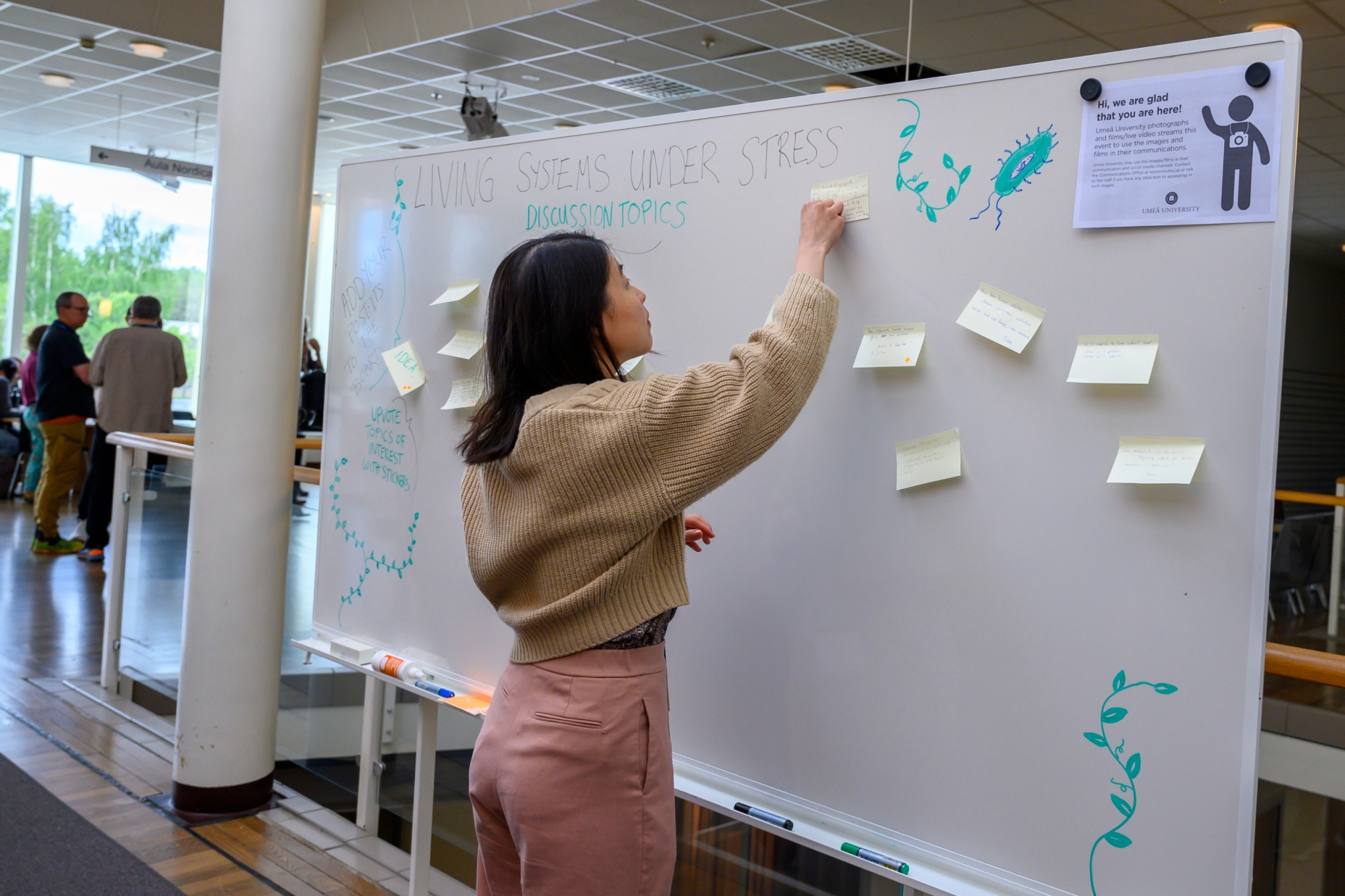
Secondary metabolite discovery and a plant wall biodesign opportunity
Secondary metabolite discovery and a plant wall biodesign opportunity May 25, 2023 Watch Lunch Pitch Video Pitch 1: Laura Carroll: Machine learning approaches for microbial [...]
Predicting gene expression, microproteins, and new tools
Predicting gene expression, microproteins, and new tools May 11, 2023 Watch Lunch Pitch Video Pitch 1: Jian-Feng Mao: Predicting allele-specific gene expression (ASE) in poplar [...]
Algal photosynthesis, statistical tools, becoming Bayesian, and modelling cell shape.
In search of collaborators to study algal photosynthesis, becoming Bayesian, cell shape and apply statistical tools. May 4, 2023 Five researchers pitched projects during the IceLab multidisciplinary postdoctoral project call information [...]
Conifers adapting to light and using autoencoders on zero-filled data
Conifers adapting to light and using autoencoders on zero-filled data April 27, 2023 Watch Lunch Pitch Video Pitch 1: Sonali Ranade: Adaptation in the Christmas [...]
Metabolism: not the limiting factor in prokaryotic endosymbiosis
Metabolism: not the limiting factor in prokaryotic endosymbiosis “One of the great mysteries of biology,” says Eric Libby, former SFI Postdoctoral Fellow, now an associate professor at the Integrated Science Lab (IceLab), Umeå University in [...]
In memory of Carl Kempe
In memory of Carl Kempe Permanent secretary, Kempe foundations We are deeply saddened by the passing of Carl Kempe. Carl was integral in shaping our creative culture for disseminating [...]